Specialities of Homeopathy medicines


Mode of taking Homeopathy medicines by Dr S. Chidambaranathan B.H.M.S, M.D Homeopathy Doctor Madurai   (Treatment avail for all people around the world)


HOMEOPATHY – Mode of taking Homeopathy medicines


Under the diagnosis and advice of a qualified Homeopath, Homeopathy medicines will usually give comfort, calmness and hope to suffering mankind even in difficult cases, for some of which the modern medical world says nothing can be done. Homeopathic medicines can provide cure gently, promptly and permanently if they are taken properly. The medicine, its potency (power), its frequency (repetition), may vary according to the patient and the condition. Even with long time medications, Homeopathy medicines are completely safe and free from all sorts of side-effects and addiction. It can be safely used in infants, children, adolescents, adults, elderly people and even in pregnant women.


Medicine administration is not child’s play. It should be dealt with carefully by experts. One should be aware that success in treatment does not entirely depend on God or doctor or with patient cooperation/management. Everything should go hand in hand for fast cure. Finally, it depends on the patient who takes the medicines. A patient needs to take care in all aspects, in correct administration of medicines and safe handling of it to avoid contamination.
Care equals cure 


Care should always be taken in all aspects while taking Homeopathic medicine i.e., in preserving and serving the medicines, by way of administration, by way of following restrictions, etc. If one takes care of medicine handling and promptness in intake of medicines, Homeopathy will never let one down. Simply ignoring the proper way of intake is like losing track in spite of having a torch (right medicines from right doctor). Also, the “best of all the medicines” is rest, sleep, water and diet, without which in no way treatment will be successful.
Homeopathic medicines – come in different forms i.e., in pill form, tablet form, trituration powder form, dilution or liquid form, oil form, ointment form, etc. Since all Homeopathic medicines come with a base of alcohol or sugar of milk or vaseline / gel / oil, mostly the medicines taste the same: they cannot be identified or redefined once the label is removed. So, care must be taken to avoid confusion by losing the label.
The healing secrets of Homeopathic medicines are
  • Take right medicines in right dosage, from the right doctor.
  • Don’t self medicate.
  • Avoid drug cocktails, like adding a pill for each complaint or symptom.
  • Timely meals, good amount of water intake, regular exercise, good aeration, good sleep, etc., will usually support one’s healing process.
  • Ruling out causative factor and following proper diet with restriction (if any) will also lead one to be cured at the earliest.
Safety measures in handling Homeopathic medicines – Usually, costly things will be cared and stored well. Health is more than wealth, so health promoting Homeopathic medicines should be cared for more. One must know what should be done and what should not to be done.
  • Homeopathy medicines usually come in minimum dose, so any strong agent will suppress the power of Homeopathic medicines. To realise or attain full effects of Homeopathic medicines, it is better to keep them safe in a cool, dark and dry place, i.e., it is better not to keep Homeopathic medicines under direct sunlight, heat, in the midst of aromatic or strong odour substances.
  • It should be cared well for avoiding contamination
  • It should be kept out of reach of children since due to its sweet nature, children often take the entire pills at one go which may lead to complications.
  • The medicines mostly come with alcohol base, so it will evaporate if left open for a long time. So care should be taken to keep it well closed i.e. air tight.
  • While taking globules, it is better to avoid handling or counting by hand to avoid contamination. The container cap itself can be used for dispensing the medicine without touching the globules.
Expiry of Homeopathic medicines – Usually, homeopathic medicines do not come with expiry markings, since they are mostly prepared with the base of alcohol, which is a very good preservative (like old wine).
So if there is no contamination, it can be used for several years.
Determination of the way of Homeopathic medicine intake


  • Medicines should be taken with a clean tongue – Unlike other systems of medicines, Homeopathy medicines don’t act through blood after digestion of the drug but they act through nerve endings in the tongue to reach the brain and work from a higher level. So it is better to avoid taking anything (foods or drinks) 10 minutes before and after taking the medicine i.e. medicine should be taken on a clean tongue. Clean tongue means one need not wash the tongue, but simply that it should be devoid of any taste. For example – if you take coffee or tea, the taste of it will remain for 15-20 minutes. So in that period one should avoid taking medicines.
  • Medicines should be allowed to dissolve in/under the tongue on their own and are not to be swallowed. Don’t alter (increase or decrease) dosage – follow the doctor’s advice.
  • Medicines can be taken before or after food, even on an empty stomach (since it will not cause any ulceration, also it will dissolve in the tongue itself and won’t reach the stomach to cause any effect). But at the time of administration of the drug, the tongue should be tasteless and free from all hindrances.
  • Don’t touch the medicines with hand for counting the tiny globules or for taking it, since it may get contaminated. Discard contaminated medicines.
  • There is no specific restriction in food while on Homeopathic medicines. But some medicines may need specific food restriction since that type of food may have inimical action against specific medicines. But any way, it is unnecessary to follow strict diet restriction unless otherwise the situation requires or the doctor calls for it.
Restrictions 
Some would say that onion, garlic, tamarind, tomato, asafoetida, pepper, coffee, tea, etc., should be strictly avoided while taking Homeopathic medicines. Actually, it is not so always. Likewise, some Homeopaths restrict perfumes and sometimes some doctors would suggest not using even paste. But the real need for restrictions usually differs from person to person depending upon their suffering and the selection of drug. So the doctor would mention the required restrictions, if any. But anyway, alcohol, smoking, tobacco chewing and mouth fresheners should be strictly avoided since these hinder the action or acceptance of drugs.
Restriction cannot be followed in all cases, i.e. because of unavoidable situation as nature of work, surroundings, etc. For example, if one suffers from pollen allergy, is the advice for shifting of place to avoid pollens advisable? Is it feasible all the time for everyone? In such a situation, if one can avoid it, it is totally unnecessary to take medication since there won’t be any complaints thereafter.
If the patient is going to live in the same situation in future also, any type of restriction (unless it is irrevocable like in diabetes, one needs to restrict / avoid sugar, likewise in hypertension, one needs to restrict / avoid salt compulsorily) will give him only temporary relief (for example – for skin allergy many doctors recommend restricting the eating of brinjal, fish, tomato, etc.). This doesn’t mean that one can dabble in allergens / irritable agents / causative factors during treatment, but one need not go for total deprivation.
Also, following strict restriction is not that easy i.e. none could follow restriction in every aspect. Also restrictions could not be maintained all the time and in all places. Also, in these types of cases, people, after feeling better, cannot judge whether the improvement was due to the medicines or the restrictions. Here medicines cannot wholly be claimed to be the curing agent.
If the sufferer happens to take these restricted things later, they may immediately feel the complaint again, i.e., a simple variation may often bring on a flare-up. But if he is cured without any restrictions, amidst all the pollutants, by raising immunity, he will be cured once and for all. One can live with his desires and aversions because everyone is living in a polluted world where water, food (fertilisers) and air is polluted. So one cannot get away from it / should follow restriction for it. Homeopathy can comfort / ease modern (polluted?) living.
Expecting good health instantly, some patients will switch over to Homeopathy from other schools of medicine throwing off their long-term medicines. It is not safe to withdraw like this, since they would have developed dependency due to long time usage. For example conditions like BP, sugar, thyroid, epilepsy, steroids, etc. If one does like this, the worsening of complaints due to skipping of medicines will be unnecessarily blamed on Homeopathy. In this case, one has to go for tapering of medicines on getting better after taking Homeopathy medicines along with the previous one with doctor’s advice.
Homeopathic medicines can be safely taken along with allopathic medicines or other systems of medicine, but it is better to avoid them at least half-an-hour before and after taking Homeopathy medicines. Likewise, are the vitamins, minerals and health drinks that one has been taking for quite sometime.


for new hope

Dr. S. Chidambaranathan, BHMS, MD (Homeo)
Laxmi Homeo Clinic
24 E. New Mahalipatti Road
Madurai, TN 625 001
India

Tel:  +91-452-233-8833 | +91-984-319-1011 (Mob)
Fax: +91-452-233-0196
E-mail:  drcheena@yahoo.com


(Disclaimer - The contents of this column are for informational purpose only. The content is not intended to be a substitute for professional healthcare advice, diagnosis, or treatment. Always seek the advice of healthcare professional for any health problem or medical condition.)

urine facts and nature


Urinary infection and kidney by Dr S. Chidambaranathan B.H.M.S, M.D Homeopathy Doctor Madurai   (Treatment avail for all people around the world)


URINARY SYSTEMS – All about urine


Water is essential for survival of life. The water we take washes and
cleans our body and comes out as urine. Water usually is lost from our body through skin, lungs faeces and urine. Our body’s outer surface is kept clean by bathing with water and cleaning of inner organs and tissues are done by blood plasma by the process of osmosis or electrolyte transportation between tissues with our water intake. This blood plasma is filtered by kidneys and liver and kept clean and perfect. The waste product thus filtered out by the kidneys is urine.
Drinking water and pissing (process of urination) is inevitable for every animal or human being. There is a saying that “we can well control our bouts of anger, but not the urine”. Everyone might have experienced the pleasure of passing urine after controlling it for a long time. Children do pissing as an art. A newborn has to urinate within 12 hours of the birth time or otherwise it should be seriously investigated. Urination is so much important for our body to maintain it clean and make it non-toxic. The digestive tract or stool habit can go on strike (without passing stool) for days, sometimes even for a week, but our kidneys cannot take rest even for a single day.
Urine can indicate 
1. Urinary tract infection with lab analysis
2. Systemic illness with increased frequency and volume of the urine, such as renal excretory disorders, hypertension (in initial stage, by natural process, urination frequency and volume of urine is increased, so automatically blood volume as well as blood pressure gets lowered), anaemia,etc.
3. Metabolic disorders with increased frequency and volume of the urine with glucose (diabetes), increased elimination of fructose (fructosuria), uricosuria (gout), etc.
4. Pregnancy in women. It can also show adulteration in foods (food colours, presence of toxic substances, etc.), chemicals in water, etc. when analysed in a lab
5. It can also show the culprit or cheat who takes steroids in sports. Likewise, it can also help to identify persons who have ingested alcohol or drugs
Urine often cautions one about a disease in the early stages itself. If we do not take care, the disease will get rooted and expand leading to damage of the inner organs. Nowadays, everyone hears about old people or devotees or leaders or their followers or diseased people drinking their own urine to stay healthy or cure their problems. Is it needed for cleaning the bowels, as said by urine therapists? Does urine contain active ingredients or has medicinal values? What is urine? How it is formed? Why does it get eliminated? Does disease get reflected in urine? What are all the diseases or conditions that get reflected in urine? What is to be noted during urination? Someone complains about yellowish urine and someone else about hot urine. Does this denote body heat or is there any inference from it? How to test it? Why is urine analysed in a lab? We shall analyse these questions one by one.

What is urine? 
Urine is a body’s eliminating fluid which is supposed to be a waste product. It usually contains waste or excess materials from our body, toxic substances, etc. Its main constituent is water.


Normal composition of urine
 is water – 95 per cent, other major and common constituents are sodium – 0.4 per cent, ammonia – 0.05 per cent, phosphates – 0.6 per cent, urea – 2 percent, sulfate – 0.2 per cent and creatine, urobilinogen, casts, etc. in minute quantities. Normally, soluble substances are excreted in the urine.

How it is formed?


Urine is produced by the kidneys as a process of cleaning the blood and excreting the unwanted soluble waste products from the body. Millions of nephrons in the kidney filter out the waste products, toxins, excess water and mineral salts from the bloodstream. Kidney, the leader of water balance mechanism, eliminates nitrogenous wastes in a non-toxic concentrated form. To maintain fluid balance, it filters, cleans and purifies about 150-180 litres of blood and recycles it daily. The urine formed in the kidney is drained by ureter into the bladder for storage. The urethra brings out the urine from the bladder, when time and place permit. Urination is normally controlled by a reflex phenomenon with urinary sphincters.

Since kidneys work as an eliminator and absorber with their permeability, they can also control the blood volume by adjusting urine volume to control blood pressure naturally. When there is dehydration, it reabsorbs the water from distal tubules and balances the blood volume. In case of hypertension or excess fluid intake, it eliminates the water and reduces blood volume and blood pressure. So, till the urine gets into the bladder from the kidney, it has control over urine in every aspect. The kidneys maintain blood acidity by excreting excessive alkaline salts for controlling infection in the blood. The action of the kidney is mainly controlled by endocrinal glands.

Why does it get eliminated?

Urine is eliminated because it is considered a waste product or an excess product. So, nature cares for its elimination, to avoid overdose or toxicity and compels the kidney to do the elimination process i.e. elimination of urine. In order to maintain blood acidity, excess alkaline salts are filtered in the kidney. The filtered product should be excreted anyway, so with the vehicle of water medium these salts are excreted in the urine.

Is it needed for cleaning the bowels, as said by urine therapists?

No, it is not necessary. The water we take is enough for the business in the bowels.

Does urine contain active ingredients or has any medicinal value?

No. Nothing has been proved like that till now. Since urine is decided as an unnecessary waste product by nature or the body, it gets eliminated from the body by or as natural process, so we do not need to recycle it for any purpose.
People who follow urine therapy mainly claim by quoting that the amniotic fluid that surrounds babies in the womb is supposed to be primarily the urine of the embryo which has quick healing effects on abrasions or bruises or cuts or any lesion, like sea water.
Till now, with active research, no medicinal value is evaluated as such.

Do diseases get reflected in urine?


Yes, most of the diseases in the initial or final period reflect their nature in colour, volume, sp gravity and contents of the urine. Most of the diseases of the bladder or urinary tract are often faced by women than men since the male urethra is 6-8 inches long whereas the female urethra is only 1½ inches long. So any infection can easily enter into the bladder and ascend to the kidney to disturb the urine and its constituents. Also the urethra of women is very close to the vagina, so any infection of one can spread to the other.
Diseases often reflect their occurrence or presence with the following symptoms or problems:
  • Dark, scanty and strong smelling urine – in case of infection (cystitis- infection of the bladder, nephritis – infection of the kidney, urethritis – infection of the urethra, ureteritis – infection of ureter, etc.)
  • Burning urination, difficulty in urination and painful urination – infection and obstruction
  • Reduced volume (oliguria) – infection, obstruction, renal stones, kidney failure, etc.
  • Increased volume of urine (polyuria) – hypertension, diabetes, nephrotic syndrome, intake of beer or alcohol or drugs, endocrinal disorders
  • Absence of urine (anuria) – obstruction or kidney failure or stenosis
  • Sweetish smell of urine or sugar in the urine (glycosuria) – diabetes, kidney damage due to drugs, toxins, etc.
  • Offensive urine or proteins in the urine (proteinuria) – diabetes, kidney damage due to drugs, toxins, etc.
  • Bloody urine (haematuria) – infection or renal stones
  • Yellowish urine with bilirubin in the urine – liver disorder – jaundice, cirrhosis, etc.
  • Semen in urine with great exhaustion (semonorrhoea) – prostate problems
  • Backache early in the morning – infection, renal stones (due to fullness of kidney, pelvis)
  • Urinary tract infection with Leucorrhoea in females – due to spread of infection into the urinary tract (honeymoon cystitis occurs in newly-married females due to overindulgence in sex)
  • Fatigue and exhaustion – Renal problems when excreting needed constituent in urine
  • Lower abdominal pain or supra pubic pain – due to infection or stones
What are the diseases or conditions that get reflected in urine?
Diabetes, hypertension, metabolic disorders, renal disorders, liver disorders, jaundice, urinary tract infection, high fever, etc., can reflect their presence in urine. Nervous disorders like paralysis, paraplegia, cerebral lesion & cerebellar lesion can hinder bladder emptying capacity and reflect in stagnation and infection. Conditions like increased sweat, summer, air-conditioned room, heavy exercises can also reflect in urine in its concentration and volume.

What is to be noted during urination?

Force, flow, frequency, control, volume, pain, colour, odour and gravels or deposits.
Force, flow, frequency and control of urine – Always talk to a doctor and get the urine analysed, if there is any intermittent flow, uncontrolled urine, difficulty in starting the urine stream, spraying of urine during urination, lack of a force in stream of urination, dribbling urination, slow thin stream of urination, etc. The common problems affecting force and flow of the urine are urinary tract infection, urinary obstruction, urethral stricture, phimosis, renal stone, congenital abnormalities like narrow urethral opening (meatal stenosis), etc.
Frequency of the urine is often found when there is increased intake of water or in diseased condition like diabetes, hypertension, etc. Small bladder and urinary tract infection can also cause frequency of the urine due to early fullness or irritability due to infection and a disability to hold the urine for a long time. Here also, there won’t be any satisfaction after passing the urine since the weak bladder cannot constrict fully to empty the urine. The stagnated urine further irritates the bladder and produces the impulse for urination and makes the person go to the toilet often. Urinary incontinence or uncontrollability can be seen in nervous affections like paralysis or paraplegia or autism or cerebral lesion or cerebellar lesion or spinal injury causing damage to spinal nerves. To treat these types of ailments, the complaints should be evaluated in a detailed manner to attain diagnosis and treatment. Anxiety and tension in mind also causes frequent urination due to increased blood circulation.
Normal volume 


Normal volume of urine excreted is 800 ml – 2200 ml/24 hrs. It varies with the intake of the water or fluids. Urine volume normally reflects the organism hydration state. It is according to body activity or exercise, but usually ¾ th of the ingested liquid should be excreted as urine. It is mostly regulated by ADH (anti diuretic hormone). The increased amount of water intake will induce increased excretion of urine (polyuria). In case of increased water intake and increased urine elimination, the excreted urine will be very much diluted or has low concentration of solutes. In a dehydrated state or during diarrhoea or vomiting, the anti-diuretic hormone gets increased in circulation and increases the permeability of the renal collecting tibule epithelium, so that maximum amount of water is re-absorbed. Hence, urine formed in this occasion will be scanty and very much concentrated with solutes.
The volume of urine usually increases in case of increased fluid intake, diabetes, hypertension (BP) and decreased in case of reduced fluid intake, low blood pressure, nephritis, fever, diarrhoea, vomiting, shock, burns, etc. The volume of the urine mainly depends on the presence of sodium, which in turn depends on aldosterone – a mineralocorticoid hormone secreted by the adrenal glands. Sodium holds water in plasma itself, so only in case of odema or hypertension doctors often advise reduced salt intake.

Colour
 – It is normally pale yellow or amber or clear depending upon a person’s health, water intake and diet. Even an uneducated man would get suspicious if there is any change in colour of the urine. Change in colour is mainly due to bilirubin. It may also change due to drugs, colours, dyes and chemicals used in sweets, foods, etc.
  • Yellowish urine indicates presence of bilirubin (jaundice) or excess of vitamin-B
  • Cloudy turbid urine always denotes infection and loads of bacteria
  • Smoky urine indicates the presence of blood cells or any other chemicals
  • Red or brown urine indicates the presence of blood (renal stones, infection, etc.)
  • Whitish urine indicates the presence of phosphate of lime
Frothy urine is often passed in case of jaundice or renal damage. It indicates the presence of bile salts or albumin.

Odour
 – Urine mostly has the odour of ammonia, due to the nitrogenous wastes present in the urine. Aromatic foods can also affect the odour (large amount of asparagus intake can cause a putrid odour during urination). Urine may smell like sweet or fruit in case of diabetes and large amount of ketone bodies excretion. Usually, adult urine is smellier than children’s since adults take a higher protein diet. The bacteria present in the urine usually reduce nitrate to nitrite with the presence of ammonia in the urine. This gives it the smell. Normally, this bacterial action needs 4-5 hours to occur. So, morning urine or concentrated and stagnated urine is smellier than normal, frequently passed, diluted urine.

Gravels or deposits
 – are minute sediment particles present in the urine. They are noticed when the urine is much concentrated and are seen at the bottom of the sample or found like sand in the urine. They can be viewed easily after getting centrifuged in the lab. They are supposed to be coagulated protein casts, cell debris, RBCs, WBCs, bacteria, pus cells, tiny stone gravels, etc.
Somebody may often complain about yellowish urine and some others about very hot urine. What is the inference?
Actually, yellowish urine denotes its concentration. It is always seen in cases of scanty urine, especially after heavy exercises or long distance travel or sunbath. Urine is scanty and concentrated due to an alternative pathway of water elimination, i.e., through sweat, to maintain the thermostat of the body. If sweat is more, then urine will be concentrated more due to less availability of water to carry solutes. If water intake is increased, the concentration automatically gets diluted and the colour will resume to normal. But if you find yellowish urine even after plenty of water intake, it is better to rule out jaundice.
Usually, morning urine is more concentrated than at other times since we do not drinking water at night. So, to maintain the water balance of our body, less water is excreted and thus the urine becomes more concentrated.
Hot urine is a reflection of the body’s internal temperature, i.e., it is felt more during the cold weather (where the body maintains internal temperature as warm as it can) or after violent exercises (where the body temperature is high due to increased body metabolism).
How to test urine?
By urinalysis
What for it should be analysed in the lab?
Urine tests are simplest, painless, cheapest, non-invasive and highly informative, which can guide treatment. In all urinalysis, normal value varies with age, sex, time, device used, solvents or kit used, analyser, sample collecting techniques (after fasting, before fasting, etc.), etc.
Even though urinalysis is mostly done in case of a urinary tract infection and renal complaints, it is necessary to do in all cases of prolonged complaints or diseases, to rule out any systemic complication. Also, it should be made part and parcel of routine periodic health examination to analyse our body condition.
How should urine be analysed?
For analysis, urine samples must be collected in a proper way, i.e. care must be taken in collecting the sample in a clean, fresh, sterilised bottle or container without any contamination. The best urine sample is midstream of first (morning) urine.
Most of the laboratories are now doing urine test and blood test with automated analyser which gives report in printed colour strip form. They measure everything electronically.

Investigations


Microscopic examination
 – urine should always be evaluated microscopically for the presence of casts, crystals, cells and bacteria.
Lab or chemical analysis of urine – is done for evaluating pH, specific gravity, sugar, albumin, ketone bodies, bile salts, pigments, blood, etc.
pH – (hydrogen ion concentration which helps in denoting acidity or alkalinity of the solution variation from 1-14 i.e. from most acid to most alkali). Urine is usually slightly acidic i.e. below 7 (indicated by blue litmus paper turning red on getting dipped) but normal range is between 5.5- 8.0. Occasionally, it becomes alkali (red litmus paper turns blue) during infection where pH is raised.
pH of urine in case of child – 6.2 – 6.8
pH of urine in case of adult – 5.2- 8.0

Specific gravity
 of the urine changes according to the volume of the urine and solute present in the urine. In case of reduced volume of the urine in normal excreting solutes, the sp gravity is raised; whereas in increased volume of urine with normal excreting solutes, it is lowered. Increased level of protein in the urine also elevates the sp gravity.
Humans are soft-skinned persons who sweat more to produce more hypertonic (highly concentrated) urine. Insects, spiders, reptiles, birds and other animals excrete less hypertonic urine since they have hard body protective surface or hair or feathers which usually protect loss of fluids from sweat. Fish and other water animals always pass hypotonic (less density) urine. Also, humans take a good amount of sodium salt in diet which will hold or retain water content in the body. The distal renal tibules in humans are also designed in such a way to reabsorb most of the water and salts to maintain our body water balance which causes urine to be more hypertonic.
Specific gravity normally ranges from – 1.001 – 1.035
Normal sp gravity of urine in children – 1.002
Normal sp gravity of urine in adult – 1.030

Sugar or glucose in urine
 – Glucose is always absorbed more

rapidly than water. If there is excess glucose in the blood due to lack of insulin (which is essential for glucose metabolism in the body) or when there is kidney damage due to drugs or toxins, glucose can occur in urine (glycosuria). In this case, urine test for glucose should be done in 12 hours fasting time and also, after two hours of food intake (post prandial) to analyse it thoroughly. The presence of sugar is now analysed by two methods, i.e., one by old Benedict’s reagents and another by latest dipsticks. In case of Benedict’s test, if results shows
Blue colour – Nil
Green colour – + (0.1- 0.5 g/dl)
Yellow colour – ++ (0.5- 1.0 g/dl)
Orange colour – +++ (1.0- 1.5 g/dl)
Brick red colour – ++++ (1.5- 2.0 g/dl)

Protein in urine
 (proteinuria) – Acceptable normal excretion range of protein in urine is below 0.15 gms/24 hours of urine. This occurs usually due to day-to-day activities or exercises where protein metabolism is used for energy. Proteins occur in urine in diseases like kidney disorders (nephrotic syndrome, renal failure, pyelonephritis, etc.) heart failure, gout, malignant hypertension, toxicity of high fever, drugs, eclampsia (convulsive disease due to high BP and proteinuria in pregnant women), etc. They usually appear as casts in urine, which can be confirmed by their coagulation or white precipitation while heating.

Albumin
 – Normal range of albumin in urine is <3.5mg/mmol. Test for Microalbuminuria (increased elimination of albumin) is most important test to be done in diabetic patients to rule out kidney involvement or damage. Since albumin level varies with day-to-day activities and exercises, it is better to test after overnight rest. It is often traced in cases of heart failure, renal failure, hypertension, urinary tract infection, etc.

Urea
 – is the end product of protein metabolism. A high protein diet will increase urea level (normal 20-40 mg /100ml) in blood and urine. It will be abnormally high in cases of kidney disorders (where there is defect in filtration and elimination) and diabetes (due to use of protein metabolism for energy since glucose metabolism is impaired). Normal acceptable range of urea in urine is 180-600mmol/24 hours.

Ketone bodies (Acetone)
 – normal level in urine is <500mg/24 hours – starvation and uncontrolled diabetes can elevate the levels.

Bile salts and bile pigments
 – give yellow colour to urine when they occur during jaundice

Urobilinogen
 – is excreted in increased amount in case of liver disorders.
Other normal levels
Sodium – 100 -200mmol / 24 hours
Potassium – 25 – 100mmol / 24 hours
Calcium level – 0 – 300 mg / 24 hours
Phosphates – 15 -50mmol / 24 hours
Urate 1.2 -3.0mmol / 24 hours
Copper – <.6micromol / 24 hours
Pus cell and bacteria should be absent in urine. Their presence always indicates infection (pyelonephritis, urethratitis, uretitis, cystitis, etc.). If there are pus cells and bacteria, culture must be done to rule out infection. Pus cells can be easily identified after centrifuge and sedimentation.

Urine culture
 – Normally, kidney-filtered or bladder-stored urine is sterile. The small number of bacteria present in the urine while passed is due to contamination of bacterial flora in the urethra. If the result denotes more than 10,000 colonies of bacteria, then it is said to be confirmed bacterial infection or otherwise it may be due to urethral flora contamination. The common bacteria which infect are Coliforms, Enterococci, Gram negative rods, etc.
Bacteria will not sediment by centrifuge, so to identify it, culture or growth of bacteria in a medium is usually followed which can easily show the type of bacterial infection. Nowadays culture tests are done with sensitivity tests, so that the correct drug can be administered in the initial period itself.

Casts
 are coagulated proteins seen as sediments in urine, so proteinuria is the precursor of cast formation. The blood cells (RBCs & WBCs) and epithelial cells can get lodged to this and form erythrocyte casts, leucocyte casts and epithelial casts. In normal conditions, casts will not be seen in urine.
Hyaline casts – are protein casts which occurs usually after violent exercises and also in case of chronic kidney failure
Leucocytes casts occur after urinary tract infection
Erythrocyte casts occur in hypertension, anaemia, renal stones, urinary tract infection etc.
Epithelial casts show inflammation and degeneration of kidney

Crystal
 formation occurs in the urine when it gets supersaturated with the solutes. It can produce uroliathiasis: for example – stone formation from increased excretion of uric acid which may sediment and form crystals or stones. Likewise are the cystines and oxalates.
Round, brick red/orange sediment in the urine of children indicates Xanthine.
Pink or yellow colour crystals – cystines
Smooth and chalky – phosphates
Dark brown – oxalates
Red urine – The high intake of beetroot usually resemble blood in the urine; another factor which can mimic bloody urine is dyes and chemicals used in sweets. In diseased conditions, bloody urine (Haematuria) may occur due to infection or stones in kidney or bladder or ureter or urethra or due to infection of the prostate. Everyone should be ever careful if blood cells are present during urination since it indicates that the condition is serious. Most of the time, it is passed as microscopic haematuria (i.e. blood in the urine may not be visualised normally). But sometimes, in case of massive bleeding, urine appears as bloody or pinkish or smoky or deep brown, alarming the person. Even though we might pass 1-3 RBCs (red blood cells), elevated levels will usually indicate internal diseases or complications. The RBCs occur in the urine in abnormal conditions like kidney stones, cancer of the kidney or bladder or prostate, heavy urinary tract infection, sickle cell anaemia, etc.

Gravindex test for pregnancy
 – following a missed period, even within a week or so, pregnancy can be confirmed by detecting the presence of HCG (human chorionic gonadotrophin) in the urine. This lab test is now simplified by colour card test where everyone can use to test urine for pregnancy at home itself.
Even though urinalysis indicates a problem, finally to evaluate and analyse the effects and complication of the disease imaging studies like ultrasound scan, IVP, CT scan and MRI may be necessary to confirm and proceed to the treatment.


for new hope

Dr. S. Chidambaranathan, BHMS, MD (Homeo)
Laxmi Homeo Clinic
24 E. New Mahalipatti Road
Madurai, TN 625 001
India

Tel:  +91-452-233-8833 | +91-984-319-1011 (Mob)
Fax: +91-452-233-0196
E-mail:  drcheena@yahoo.com


(Disclaimer - The contents of this column are for informational purpose only. The content is not intended to be a substitute for professional healthcare advice, diagnosis, or treatment. Always seek the advice of healthcare professional for any health problem or medical condition.)

Kidney Stones treatment and remedies


kidney Stones by Dr S. Chidambaranathan B.H.M.S, M.D Homeopathy Doctor Madurai   (Treatment avail for all people around the world)


URINARY SYSTEMS – Kidney stones
In the era of advanced ultrasound scanning, most renal stones (kidney stones) are diagnosed
without any symptom. Nowadays renal stone disease is a significant health problem. Formation of stones in the urinary tract is called urolithiasis and in kidney is termed nephrolithiasis. This causes an acute painful condition called renal colic. It strikes without warning at any time and the pain is often described as worse than labour pain.
Incidence – Men are affected by renal stones more commonly than women. The male-to-female ratio is approximately 3:1. It occurs commonly between 30-50 years of age. It is mostly found in persons whose diet is low in vitamins. Approximately 80-85 per cent of stones pass spontaneously without any mortality. Its recurrence is likely to occur if it is left without treatment.
Causes and pre-disposing factors – Stones occur due to abnormal and excessive accumulation of substances such as calcium, oxalate, uric acid and cystine in the urine. Usually, these substances are dissolved in the urine by the presence of substances like citrate and pyrophosphates. The formation of stones (renal calculi) is a complex process and depends on the various factors which include:
1) Changes in urinary PH in diseases like renal sepsis, renal ischemia and renal tumours
2) Increased urinary concentration due to dehydration – Most
stones arise in the kidney when urine becomes supersaturated with salts and crystals that are capable of forming stones. Factors of crystallisation play a major role in stone formation – like citrate, magnesium and pyrophosphate.
3) Urinary inadequate drainage – slow obstructive urine flow will encourage salt precipitation due to stagnation
4) Hyperparathyroidism may also lead to hyper calcuria (increased calcium in urine)
5) Vitamin A deficiency
6) Increased intake of vitamin D, calcium, protein, tea or fruit juice
7) Prolonged immobility where stone formation is due to hypercalcuria
 Metabolic disorder (e.g., hypercalcemia or hyperparathyroidism)
9) Hereditary disease (e.g., cystinuria)
10) Underlying disease of the small bowel
11) Urinary tract infection
Symptoms: 


Many stones are asymptomatic until they begin to move down the ureter, causing pain due to obstruction. The main symptoms of renal colic are:
1) Pain – The most common symptom of kidney stone is severe back or abdominal pain. The colicky-type of pain is the sudden onset of very severe pain sub costally and it radiates interiorly and anteriorly towards the groin. The pain generated by renal colic is primarily caused by the dilation, stretching and spasm caused by the acute ureteral obstruction. As the stone moves lower, the pain may be felt in the genitals, especially the testicles in men and the labia in women. In infants, stone pain is often confused with colicky abdominal pain. Most of the calculi are originating within the kidney and proceed distally, creating various degrees of urinary obstruction as they become lodged in narrow areas, including the ureteropelvic junction, pelvic brim, and ureterovesical junction. Location and quality of pain are related to the position of the stone within the urinary tract. The severity of the pain depends on the degree and site of the obstruction and not on the size of the stone. Pain also varies in intensity with the presence of ureteral spasm and infection
a) If stones block in the ureteropelvic junction – pain will be severe with a deep back pain without radiation to the groin and due to distension of the renal capsule.
b) If stones block the upper urethra and renal pelvis – Pain will radiate from back to frontmisguiding to gall stones on right and peptic ulcer in left.
c) If stones block the middle urethra – pain will radiate anteriorly and
suprapubically. This mid urethral pain usually misguides to appendicitis on the right.
d) If stones block the distal urethra – pain will radiate into the groin or testicle in the male or labia majora in the female. If a stone is lodged in the intramural urethra, symptoms may appear similar to cystitis or urethritis. These symptoms include suprapubic pain, urinary frequency, urgency, dysuria, stranguria, and pain at the tip of the penis.
e) If stones enter the bladder, pain is often switched off and stone gets circular or oval shape as it rolls inside the bladder and is passed easily during urination.
2) Nausea and vomiting with excessive sweating – This is due to the common nerve supply by vagus nerve to renal pelvis and bowels. This may also misguide the patients that it these are bowel disorders.
3) Urinary tract infection – Symptoms may include fever, chills, sweats and pain with urination. Kidney stones and urinary tract infection can cause block with urine retentions. Red blood cells and pus cells will be found in the urine. Also albumin, hyaline casts and crystals will be seen.
4) Fever – It is not a part of the presentation of uncomplicated nephrolithiasis. If fever is present, rule out hydronephrosis, pyonephrosis, or perinephric abscess. Such a condition is potentially life-threatening and should be treated as a surgical emergency.
Stone analysis – Stones which are passed spontaneously should be saved for analysis of its type and composition. Chemical analysis of a calculus passed in the urine or removed surgically is very helpful in identifying the underlying causative factor and aid in getting relief from recurrences. But in case of lithotripsy, renal stones get fragmented, and a whole stone is not available for analysis. According to composition, stone types are:
Calcium oxalate: 75 per cent
Calcium phosphate: 15 per cent
Uric acid: 8 per cent
Other types are rare which include Cystine: 1 per cent and Xanthine: 1 per cent
Stones can also be classified:


Primary stones are stones which are formed through the tendencies. For example-oxalate and uric acid and urate stones. Secondary stones are stones that occur after infection. For ex-formation of magnesium, ammonium and phosphate stones. These stones are formed in the alkaline urine. The stones formed due to vitamin A deficiency also come under this category.
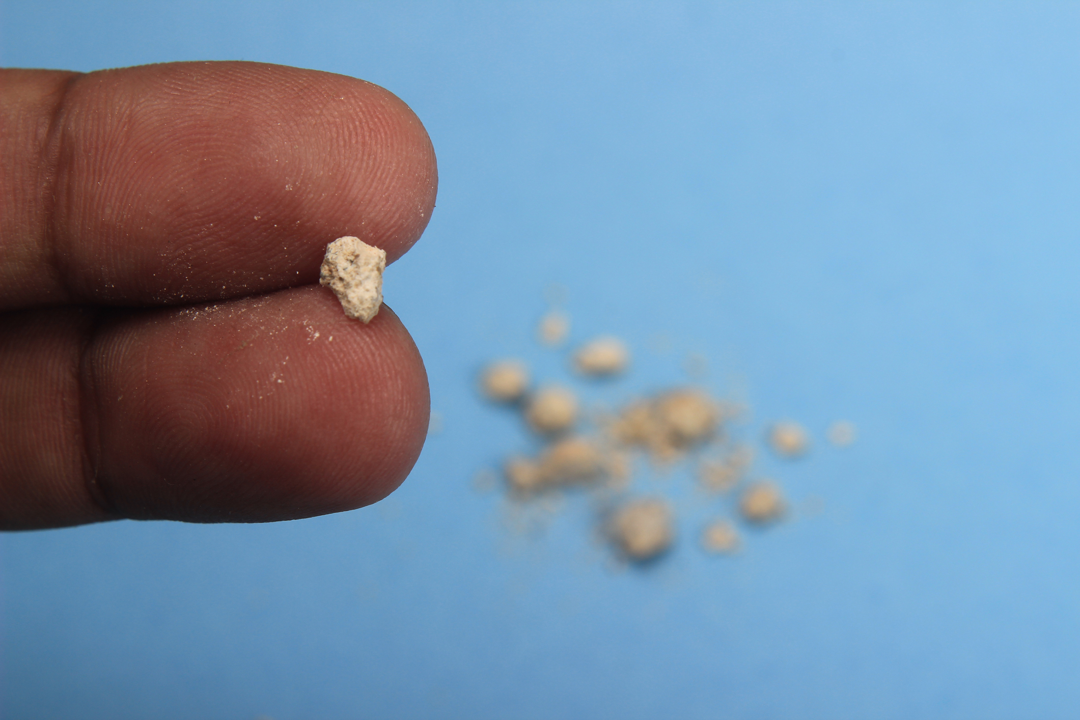
Calcium stones – Of the four main types of urinary stones, calcium stones are the most common. The calcium may combine with other substances such as oxalate, phosphate or carbonate to form the stone. Any increased levels of calcium and oxalates will increase the tendency to stone formation. Small intestine disorders and defective kidney disorders may allow too much calcium in the urine. The risk of calcium stone formation is increased in condition of hyperparathyroidism and metabolic disorders. This type of stone is extremely hard with surface rough with spicules. It is dark in colour, usually single and radio-opaque. It usually arises in acid urine.
Oxalate stones – The toxicity of oxalate in humans results from the extreme insolubility of its calcium salt or calcium oxalate, which may precipitate in the renal parenchyma or renal tract, causing either nephrocalcinosis or stones. Diseases of the small intestine increase the tendency to form calcium oxalate stones. These stones are dark brown in colour.
Uric acid stones – It is the second most common type of stone. Uric acid stones occur because of too much uric acid in the urine. They might occur due to dehydration after strenuous exercise or during an illness. Uric acid stones are common in people who have got a disease that causes high uric acid levels in the blood. For example gout patients often suffer from uric acid stones. Reduced urine volume with dehydration, hyperuricaemia, and a urinary PH that is consistently less than 6  are the important factors that influence uric acid stone formation. These types of stones are usually multiple, moderately hard, easily broken and not radio-opaque. They are brownish white in colour and usually occur in acid urine.
Phosphates stones – These types of stones are smooth and chalky with tendency to easily break. It is dirty white in colour and radio-opaque. It usually arises in alkaline urine which is favourable for infectious diseases. In this alkaline condition it grows enormously and takes the form of pelvic calyceal system assuming the shape of stag-horn. Stones are frequently diagnosed in children and women who have recurrent urinary tract infection. They can grow very large and obstruct the kidney, urethra, or bladder. A urine PH greater than 7 suggests presence of urea-splitting organisms, such as Proteus, Pseudomonas, or Klebsiella species, and struvite stone.
Cystine stones – This is a rare type of kidney stone that occurs in children of all ages due to genetic condition. Urine becomes supersaturated with cystine resulting in crystal deposition. All cystine calculi are radio-opaque. Cystine stones are pink or yellow in colour and are soft stones.
Xanthine stones occur in the rare condition of Xanthinuria which is also an inherited genetic condition. Xanthine is usually converted into uric acid. So, its diagnosis is made by low-level of uric acid. It is smooth, round, brick red/orange sediment in the urine of children.
Investigations:
Urinalysis will identify PH, and microscopy will identify cystine crystals, haematuria, pyuria and bacteria. Also culture and sensitivity tests of urine will guide on the treatment to be followed. A 24-hour urine collection is necessary in evaluating renal stones. Microscopic haematuria is present in over 90 per cent of cases with stones.
Kidney ultrasound – Ultrasound is more sensitive in detecting nephrocalcinosis and may detect radiolucent stones. Ultrasound cannot be used to find small stones (less than 3 mm).
Abdominal X-rays – Small stones may not be detected by this method and plain abdominal radiography should be performed if the suspicion of stones is high.
Intravenous pyelogram – Various reactions have been attributed to intravenous contrast agents including nausea, vomiting, urticaria, bronchospasm and cardiac arrhythmias.
Abdominal CT scan and abdominal/kidney MRI will also give a clear picture about the disease condition.
Complications:


1) Infection
2) Kidney damage – affected kidney will become functionless with hydronephrosis.
3) Recurrence of stones
Preventive measures:
Prevention with dietary changes
Drink – Plenty of water should be taken – at least 3 litres of water or an intake to make a volume of 2 litres of urine a day should be taken. After doing strenuous exercises or when passing yellowish urine, additional water should be administered. Taking tender coconut daily is good for the kidney function and also prevents stone formation.
Eat – Low-fat diet with increased natural foods and plenty of fruits should be taken. Good protein diet from vegetarian sources, such as beans is a good intake.
Avoid:
Consumption of animal protein from meat, dairy, cheese, poultry, or fish should be avoided in the case of patients having calcium stones.
Calcium supplements should be avoided.
Ice creams, chocolates, tea, soft drinks and drinks which are rich in calcium should be avoided.
Consumption of spinach, greens, vegetables, black pepper, almonds and peanuts should be avoided for patients suffering from oxalate stones.
Citrus fruits, apples, tomato, orange, lemon and strawberries should be avoided in the case of patients suffering from uric acid stones but for patients with calcium stones, it is allowed.
A sedentary lifestyle should be followed since physical activity, especially walking and jogging, can bring the stone down.

Restrict
 – Salt should be restricted
Prevention with controlling urinary tract infections

Long intervals between urinating should be avoided.
More water intake should be made.
The bladder should be emptied after intercourse as a preventive measure.

Treatment
 – Treatment goals include relief of symptoms and prevention of further symptoms. Treatment varies depending on the type of stone and the extent of symptoms and complications. Also, it
1) Depends on the size – Most of the small stones with relatively mild hydronephrosis can be treated with observation and medicines. If the stone is 4 mm or smaller, the stone is easily passed in 90 per cent of the cases. Stones of 5-7 mm have a 50 per cent chance of passing spontaneously. Calculi larger than 7 mm are unlikely to pass unassisted. In general, smaller stones are more likely to pass spontaneously. But a stone may take days or weeks or even months to pass.
2) Location of the stone – Passing of the stone also depends on the exact shape and location of the stone and the specific anatomy of the upper urinary tract in that particular individual. For example, the presence of an ureteropelvic junction (UPJ) obstruction or a ureteral stricture could make it difficult or impossible for even very small stones to pass.

3) Number of stones blocking the flow of urine out of the kidney


4) Whether it is involved on one or both sides
5) Infection – In the acute phase, intermittent pain usually lasts for 1 day to 2 days. The urinary tract does not resolve until the stone is removed entirely.
Medication – In Allopathy, the first line of treatment for a renal colic patient starts with obtaining intravenous (IV) fluids, analgesic, and antiemetic medications since many of these patients are dehydrated from nausea and vomiting.
Also most of the patients are prescribed Ayurvedic or Siddha preparation such as calcurin or cystone, etc. But getting recurrence of stones once in a year or two is very common.
Surgical option – If the stone is not passed spontaneously, surgical removal of the stone may be required. Lithotripsy may be an alternative to surgery. In this extra-corporeal shockwave lithotripsy, ultrasonic waves or shockwaves are used to break up stones so that they may be expelled in the urine or removed with an endoscope. The stones, once broken, pass out as fine sand particles. Stones are also removed using cystoscopy or ureteoscopy or percutaneous surgery.
In the case of large stones impacted in the kidneys, open surgery may be required – Pyelolithotomy – to remove stones in pelvis of kidney; Nephrolithotomy – to remove large stones in renal parenchyma without opening pelvis; Ureterolithotomy – to remove stones in the ureter that are not amenable to other treatment; and Nephrectomy – partial or the whole kidney is removed depending upon the extent of damage.

Prognosis
 – Kidney stones are usually excreted without causing permanent damage. But they tend to recur, especially if the underlying cause is not found and treated. Prognosis depends on the extent of the damage caused to the kidney. If it is involved in a single kidney, the prognosis is good.
Homoeopathic approach: 


The first medicine is water, without this even right medicine cannot work.
The second one is surely Homoeopathy medicine which can stop the tendency to form stones.
When patients suffer constantly from certain disease conditions which are untreated or not properly treated, then they become tendencies. Mostly patients who suffer from renal stones will constantly suffer from its tendencies. These types of patients can be cured permanently in Homoeopathy. Surgery can be avoided in many cases of kidney stones but in advanced conditions, surgery is the only way.
By acting in a natural way, Homeopathy increases the immune power and also removes tendencies and thus helps mankind to live in a modern polluted world.
Tricks and tips to expel stone
1) Take fluids in abundance: Drink lots of liquids so that kidney stones usually pass on their own. It increases the speed of the movement of a stone through the urinary tract. If nausea and vomiting is there, IV fluids can be given. Usually, when IV fluids are administered (3 pints) at a fast rate, it accounts for 1,350 ml of fluid. But drinking one litre of water at a time is well and good. The principle is to take a lot of fluids at a time to flush the stone out.
2) Pass the urine forcibly and speedily after getting the bladder filled up so that the stone in the pathway may get down faster with suction force and turbulence.
The early you treat, you get speedier and complete cure. In all natural way of healing, the immune power is increased against diseases so that recurrences are avoided. When the cure is not in the proper way, the disease often recurs. There are many drugs in Homoeopathy which relieve pain, to expel stones, to break stones, and for its associated complaints. Some of those are Beri beri vulgaris, Ocimum canum, Lycopodium, Sarasaparilla, Pareira Brava, Calc carb, Urtica urens, Nat phos, Terebinth, Belladona, Dioscorea, Cantharis, Hydrangea, Millefolium, Uva ursi etc., Depending upon the symptoms present in the patient, drug selection differs.
These Homoeopathy medicines not only relieve pain but also treat the condition permanently. The homoeopathic approach is a natural way to help the body by supporting the body’s own process of healing. In recent days, people are aware of Homeopathy’s miraculous cure which is permanent and safe. This is the right time for the wise to get rid of their complaints without any torture (like injections or surgeries) and live healthily


for new hope

Dr. S. Chidambaranathan, BHMS, MD (Homeo)
Laxmi Homeo Clinic
24 E. New Mahalipatti Road
Madurai, TN 625 001
India

Tel:  +91-452-233-8833 | +91-984-319-1011 (Mob)
Fax: +91-452-233-0196
E-mail:  drcheena@yahoo.com


(Disclaimer - The contents of this column are for informational purpose only. The content is not intended to be a substitute for professional healthcare advice, diagnosis, or treatment. Always seek the advice of healthcare professional for any health problem or medical condition.)

Why we are best

Best Homeopathy Treatment • We aim for Comfort First and Cure Next • You will be in safe hands experienced more than 25 years. • We can provide Best solution and Customized treatment plan for your sufferings. • You will be directly under the personal care of Chief Doctor and not with juniors. • As we had handled all sorts of cases in and abroad, we can guide you to choose effective treatment. • Further, if required to control the disease at the earliest and to be in safer side and in Incurable diseases, to improve quality of life, we can also advice you to integrate Homeopathy with Allopathy • Your treatment remains confidential with us by all means

CLICK HERE TO VIEW AND ALL DRCHEENA HEALTH ARTICLES...


Speciality Treatment

We are “The Expert” in managing chronic and incurable complaints – Here you can see a sample dozen of our area of Super Specialization. We have more proven results in following diseases

Our Specialization

Our Specialization

You can always reach us @

Laxmi Homeo Clinic CLINIC ADDRESS : 24-E, New Mahalipatti Road, Madurai – 625001 Tamilnadu, India Clinic: (+)91-452-2338833 Cell: (+)91-98431-91011 EMAIL: drcheena@gmail.com BLOG: drcheena.blogspot.com, YOUTUBE:drcheena WEBSITES: www.drcheena.in, www.drcheena.org

Disclaimer

Disclaimer: Laxmi Homeo Clinic is intending to make the people all over the world healthier through the power of information. Our health information and technologies can make a healthier tomorrow and better healthcare outcomes.
The contents of this column are for informational purpose only. The content is not intended to be a substitute for professional healthcare advice, diagnosis, or treatment. Always seek the advice of healthcare professional for any health problem or medical condition.

All rights reserved. Terms of Use